Business Inquiry
Global:
Email:marketing@medicilon.com
+1 (617) 888-9294(U.S.)
0044 7790 816 954 (Europe)
China:
Email: marketing@medicilon.com.cn
Tel: +86 (21) 5859-1500


The Medicilon translational medicine platform has an experienced professional technical team. Starting from the mechanism of action of drug targets and the clinical application of biomarkers, the team combines a variety of different technology platforms and instruments to reduce the cost and time of drug R&D, and provide diverse and efficient services for different types of drugs, different kinds of pharmaceutical companies and projects at different R&D stages.


 Summary and OutlookRelated Platforms
NanoString technology has been widely applied to the frontier fields of biopharmaceutical, including the validation of high-throughput gene expression results, gene expression profiling studies, gene regulatory network research, and clinical disease molecular classification, diagnosis and prognosis.

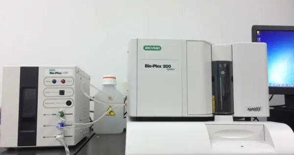
Medicilon can provide detection services for cytokines and biomarkers. It has high-end technology platforms such as flow CBA, MSD, and Luminex systems, which can detect various single plex or multiple forms of cytokines and biomarkers.

Medicilon's Bioanalysis services are compliant with the standards of FDA/NMPA GLP and involve pharmacokinetics, toxicokinetics, pharmacodynamics, immunogenicity, and bioequivalence, to support our clients.

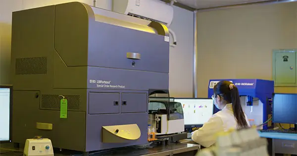
The Medicilon flow cytometry team has extensive experience in experimental operation, flow sorting method development and data analysis. The platform is equipped with a SONY flow cell sorter, which is capable of sorting of up to 6 colors at the same time, and statisfy the special scientific research and sorting needs of clients.
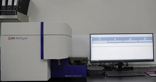
The platform is equipped with powerful flow cytometers. The newly introduced high-parameter BD FACSymphony A5 flow cytometer is equipped with five lasers and 30 parameters, enabling measurement of up to 28 colors simultaneously to meet clients' special scientific research and analysis needs.

In diagnosis, histopathological diagnosis is considered as the gold standard to determine diseases and their status. As a comprehensive CRO, Medicilon provides extensive, high quality services in histopathology.
| Paraffin Section | Frozen Slice | |
| Fixed | Before embedding: formaldehyde | Before or after sectioning: formaldehyde, methanol, ethanol, or acetone |
| Slice | Slicer | Cryostat |
| Store | Store at room temperature for many years | One year at -80 °C (longer at -190 °C)长) |
| Advantages | Easy to handle without damaging slices | ♦ Retains enzyme function and antigenicity ♦ Short protocol (usually does not require lengthy fixation steps) |
| Limitation | ♦ Overfixation can mask epitopes, increasing the need for antigen retrieval. ♦ Long processing time: Gradual dehydration in graded alcohol and xylene to facilitate paraffin penetration. | ♦ Without rapid freezing of tissue," ice crystals may form, disrupting tissue structure. ♦ Frozen sections are usually thicker than paraffin sections, which may result in lower resolution and poorer images. ♦ It may be necessary to block endogenous active enzymes. |
| Heat-induced epitope retrieval | Proteolytic enzyme-induced epitope retrieval | |
| Advantages | Retrieval of antigenic epitopes is gentler with more controllable parameters. | Suitable for antigenic epitopes that are difficult to retrieve. |
| pH value | Typically, a pH six buffer is used, but alkaline buffers are also widely used. must be determined experimentally | The pH is usually 7.4 |
| Temperature | About 95°C. | Typically 37°C |
| Incubation time | 10-20 minutes | 10-15 minutes |
| Buffer Components | Depends on the desired pH of the target antigen. Commonly used buffers include sodium citrate, EDTA, and Tris-EDTA | Neutral buffer for enzymes such as pepsin, proteinase K, or trypsin |
| Precautions | Microwave heating may result in uneven antigen retrieval. Vigorous boiling can cause debonding (separation of tissue from slide). | Enzyme repair sometimes disrupts section morphology - concentration and time need to be optimized |

 FAQs
FAQsFlow Cytometry (FCM) is a technology that allows for rapid, accurate, objective, and simultaneous analysis and quantification of multiple physical and biological characteristics of individual cells in rapid linear flow conditions. It also enables sorting of specific cell populations.
A flow cytometer is a new high-tech instrument integrating laser technology, electronic physics, photodetection technology, electronic computer technology, cell fluorescence chemical technology and monoclonal antibody technology.
Flow Cytometer is a new high-tech instrument integrating laser technology, electronic physics technology, photoelectric measurement technology, electronic computer technology, cell fluorescence chemical technology and monoclonal antibody technology.
• Fast detection speed and ability to analyze large sample volumes
Flow cytometry distinguishes itself by analyzing a large number of cells in a very short period of time. It can measure tens of thousands of cells per second.
• Simultaneous analysis of multiple features of individual cells
By using different fluorescein-labeled monoclonal antibodies or fluorescent dyes, flow cytometry can provide multiple pieces of information about single cells.
• Capability to detect diverse types of samples
Flow cytometry can analyze various types of samples including different types of cells (such as peripheral blood, bone marrow, fine needle aspirates, lavage fluids, solid tissues, cells from suspension or adherent cultures), microorganisms, and synthetic microspheres.
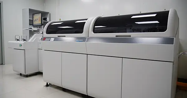
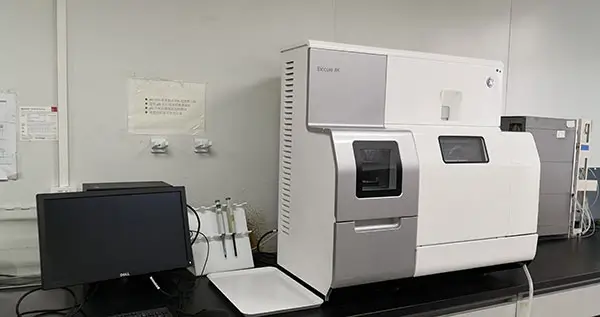
 Relevant laboratories


 marketing@medicilon.com